
The development of fluid under the retina that results in visual distortion. As the name suggests, the collection of fluid affects central vision, where it causes the most visual disturbance. Central serous chorioretinopathy is the development of fluid under the…
Just as freckles, moles, and birthmarks can occur on your skin, a nevus (the medical term for a birthmark) can also be found inside your eye. They are most often seen in the pigmented layer beneath the retina called the choroid.
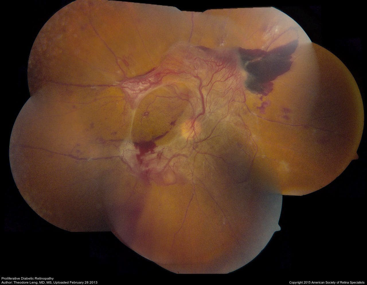
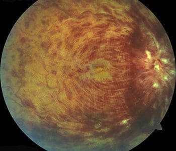
If you have diabetes mellitus, your body does not use and store sugar properly. High blood sugar levels cause damage to the small blood vessels in the retina. The damage to retinal vessels is called diabetic retinopathy. If you have…
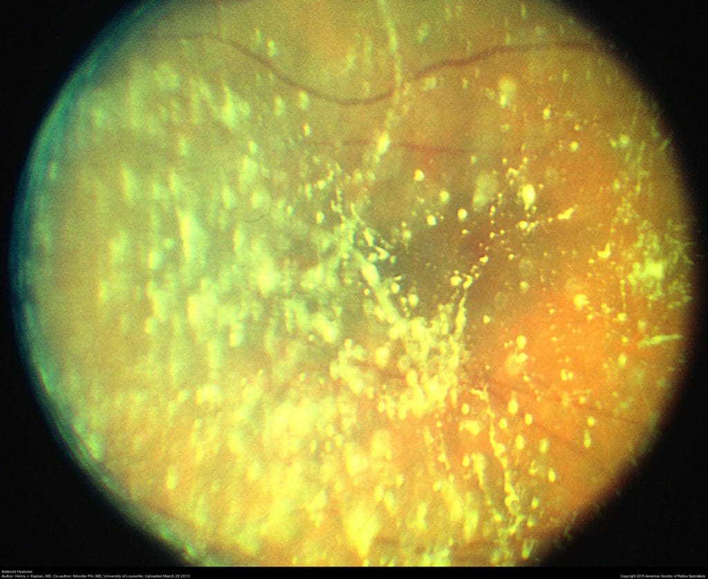
Floaters look like small specks, dots, circles, lines or cobwebs in your field of vision, while flashes can look like flashing lights or lightning streaks in your field of vision.
A disease of the human eye wherein the peripheral retina becomes atrophic in a lattice pattern and may develop tears, breaks, or holes, which may further progress to retinal detachment.
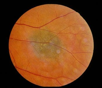
This disease is characterized by the deterioration of the central portion of the retina, which is responsible for recording the images we see and sending them via the optic nerve from the eye to the brain. The retina’s central portion…
With this condition, fluid leaks from retinal blood vessels, causing swelling or thickening of the macula. This is called macular edema and results in blurring of the central vision. https://www.youtube.com/watch?v=85nKRrcOv9I The macula is the small area at the center of…
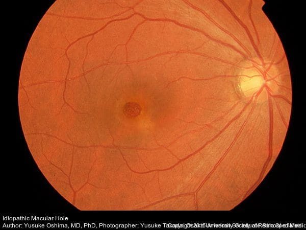
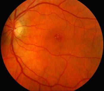
Macular holes can form when the vitreous, the gel-like substance that fills the eye and lies in front of the macula, shrinks and pulls away from the macula. In some instances, the vitreous gel sticks to the macula and cannot…
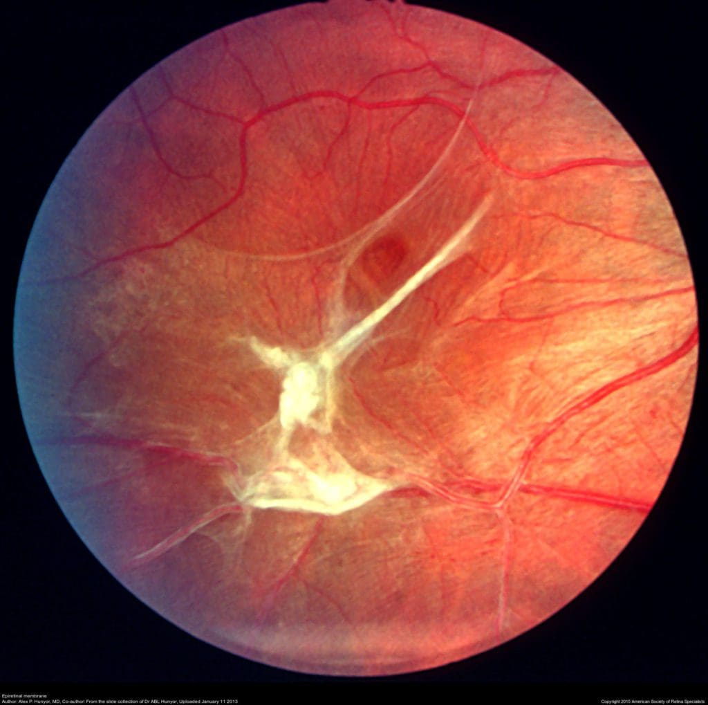
The macula normally lies flat against the back of the eye, like film lining the back of a camera. When wrinkles, creases or bulges form on the macula, this is known as a macular pucker. https://www.youtube.com/watch?v=NRWY3aIqNss The macula is the…
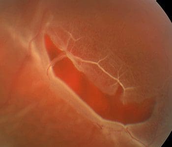
When the retina tears and pulls away from its normal position at the back of the eye, the retina is considered to be “detached.†Once pulled away, the retina no longer works properly and loses its blood supply. Retinal tears…
Retinoschisis is a disease of the nerve tissue in the eye. The retina, which consists of multiple layers of interconnected nerve and pigment cells, splits into separate layers, resulting in a loss of vision in the corresponding visual field. Retinoschisis…
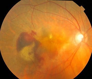
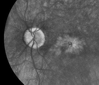
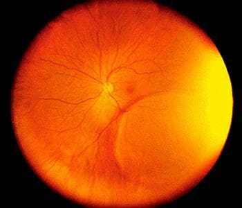
A retinal vein occlusion occurs when a vein in the retina of the eye is blocked. A blocked vein damages the blood vessels of the retina, causing bleeding and leakage of fluid from the areas of blocked blood vessels. https://www.youtube.com/watch?v=yhBbV-UoGEshttps://www.youtube.com/watch?v=jBUaNtgAaUc…
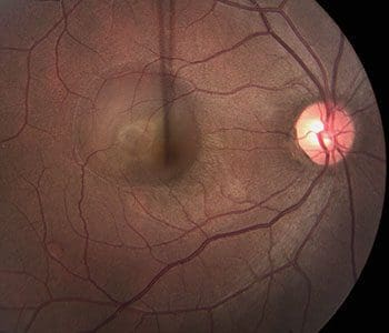
With age, the vitreous separates from the back of the eye. This separation is a natural occurrence, and for most people, happens without complication. Sometimes, the vitreous does not completely separate from the back of the eye and remains attached…
Retina Health Series
The American Society of Retina Specialists Retina Health Series offers a collection of fact sheets that offer information about causes, symptoms, risk factors, diagnostic testing, treatment, and prognoses of an array of retinal conditions.
- Age-Related Macular Degeneration
- Branch Retinal Vein Occlusion
- Central Retinal Vein Occlusion
- Central Serous Chorioretinopathy
- Choroidal Detachment
- Complex Retinal Detachment
- Congenital X-linked Retinoschisis
- Diabetic Retinopathy
- Endophthalmitis
- Epiretinal Membranes
- FEVR
- Idiopathic Juxtafoveal Telangiectasis
- Infectious Retinitis
- Intraocular Lens Dislocation
- Intravitreal Injections
- Lattice Degeneration
- LCA
- Macular Edema
- Persistent Fetal Vasculature
- POHS
- Polypoidal Choroidal Vasculopathy
- PVD
- Retained Lens Fragments
- Retinal Artery Occlusion
- Retinal Detachment
- Retinal Tears
- Retinitis Pigmentosa and Retinal Prosthesis
- Retinoblastoma
- Retinopathy
- Victrectomy
- Vitrectomy-for-floaters
- Vitreomacular-traction-syndrome