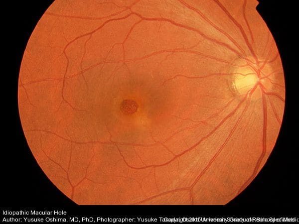
Macular holes can form when the vitreous, the gel-like substance that fills the eye and lies in front of the macula, shrinks and pulls away from the macula. In some instances, the vitreous gel sticks to the macula and cannot pull away, stretching the macular tissue, and ultimately tearing.
https://www.youtube.com/watch?v=c7vhRDuF92k
The macula is the central area of the retina, which is a thin layer of light-sensitive tissue that lines the back of the eye. Light rays focus on the retina, where they are converted into electrical signals that are transmitted to the brain and interpreted as the images you see. The macula is the portion of the retina responsible for fine, detailed vision.
Macular holes can form when the vitreous, the gel-like substance that fills the eye and lies in front of the macula, shrinks and pulls away from the macula. This occurs naturally as you age, and usually has no negative effect on your sight. However, in some instances, the vitreous gel sticks to the macula and cannot pull away. The macular tissue stretches, and after a period of time, tears and forms a hole.
Symptoms of a macular hole:
In the early stages of the formation of a macular hole, your central vision becomes blurred and distorted. If the hole worsens or progresses, a blind spot will develop in the central vision and impair the ability to see at both close range and at a distance.
If the macula is damaged, you will not lose your vision in its entirety. You will still have peripheral (side) vision.
The doctor diagnoses macular holes by looking inside of your eye. He may will also use an OCT test – optical coherence tomography – to scan and examine the retina. This test uses light waves to reveal specific layers of the retina.
Treatment of Macular Holes:
Vitrectomy surgery is the treatment to repair a macular hole and improve vision. The doctor uses tiny instruments to remove the vitreous gel that is pulling on the macula. The eye is then filled with a special gas bubble to help close the macular hole and hold it in place while it heals.
You must maintain a constant face-down position after surgery to keep the gas bubble in contact with the macula, helping to close the hole permanently. Face-down positioning is critical to the success of the surgery. Without proper face-down positioning, the chances of closing the hole and improving your vision are reduced. Proper face-down positioning is usually required for 5-7 3 days, depending upon the nature of the macular hole and the doctor’s recommendation.
will improve. The vision will improve gradually over several months to a year. The amount of visual improvement is usually determined by the size of the macular hole and its duration. The larger the hole and the longer
Learn more about Face-Down Positional Therapy.
With proper face-down positioning, closure of the macular hole occurs in over 90% of patients. With closure of the hole, the patient’s vision will improve. The vision will improve gradually over several months to a year. The amount of visual improvement is usually determined by the size of the macular hole and its duration. The larger the hole and the longer it has been present, the less visual improvement will occur. While not an emergency, macular hole surgery should usually be performed within 1-2 weeks of diagnosis.
Their are two main complications following vitrectomy surgery. Postoperative infection occurs rarely (1 in 3000 cases), while retinal detachment occurs in approximately 1% of vitrectomy surgeries. Both conditions can be treated successfully, but are sight threatening. If the patient is over 50 years old, cataract formation will be accelerated and cataract surgery will be required within 6-12 months.
The doctor will discuss the potential risks and benefits of a Vitrectomy with you before you make a decision regarding treatment.
