
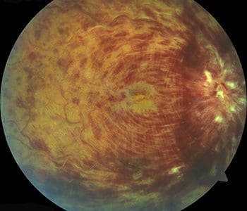
A retinal vein occlusion occurs when a vein in the retina of the eye is blocked. A blocked vein damages the blood vessels of the retina, causing bleeding and leakage of fluid from the areas of blocked blood vessels.
https://www.youtube.com/watch?v=yhBbV-UoGEs
https://www.youtube.com/watch?v=jBUaNtgAaUc
A retinal vein occlusion occurs when a vein in the retina is blocked. A blocked vein damages the blood vessels of the retina, causing bleeding and leakage of fluid from the areas of blocked blood vessels.
There are two different types of retinal vein occlusion:
- Central retinal vein occlusion (CRVO): the main vein of the eye, found in the optic nerve, becomes blockedÂ
- Branch retinal vein occlusion (BRVO): one of the smaller branches of vessels attached to the main vein becomes blockedÂ
Who is at risk for a retinal vein occlusion?
Certain illness can increase your risk for developing retinal vein occlusion, including:
- High blood pressure (most commonly)
- Diabetes
- Glaucoma
- High blood pressure
- Age-related vascular disease
- Blood disorders
If a retinal vein occlusion occurs in one eye, there is an increased chance (approximately 10% increase) that a branch or central vein occlusion will occur in the other eye in the future.
What are the complications and symptoms of retinal vein occlusion?
Macular Edema
If blood and fluid leak into the macula, swelling of the macula occurs (macular edema). Macular edema causes blurred vision, decreased vision or both.
Abnormal blood vessel growth (neovascularization)
Retinal vein occlusion can cause abnormal vessels to begin to grow on the retina. These new vessels are very fragile and may bleed or leak fluid into the vitreous. Small spots or clouds in your field of vision called floaters may appear. In more advanced cases, the neovascularization may cause the retina to detach from the back of the eye.
Pain in the eye
In severe cases of CRVO, a blocked vein causes abnormal blood vessel growth on the iris and drainage channels in the front of the eye, leading to a painful pressure increase in the eye (neovascular glaucoma). If complications from retinal vein occlusion are not treated, irreversible blindness may occur.
How is retinal vein occlusion detected?
The doctor detects retinal vein occlusions with a dilated eye examination. The doctor may perform fluorescein angiography. This involves high resolution digital photography to detect the location and extent of the blocked retinal vein and the amount of damage to the retina.
The doctor will also utilize optical coherence tomography (OCT), where he takes special photographs of the eye, to detect the presence and/or extent of macular edema.
How is retinal vein occlusion treated?
Very effective treatments have been developed recently that return vision to many patients. Anti-VEGF and steroid medications are injected into the eye to greatly reduce the swelling and bleeding that occurs in this condition. These treatments, often used in conjunction with laser photocoagulation, can reduce vision loss. Maintenance therapy is usually required following initial treatment.
Medical Treatment:
Anti-VEGF Therapy
Vascular endothelial growth factor (VEGF) is the chemical released by the retina in response to a decrease in blood supply. It promotes abnormal blood vessel growth and stimulates leakage into the retina. Injections of anti-VEGF medication in the eye can stop bleeding in the retina, stunt the growth of new blood vessels and reduce the chance of subsequent neovascular glaucoma. The therapy can also reduce swelling in the macula associated with macular edema. In some cases, anti-VEGF therapy is performed in conjunction with laser photocoagulation as discussed below.
Often, patients experience anxiety regarding the injection itself. With appropriate topical anesthesia, the injection itself is painless. The vast majority of patients are not even aware that they have had an injection. There is occasionally some discomfort after the injection due to the sterilizing solution placed in the eye to prevent infection. The doctor recommends aggressive lubrication with artificial tears or ointment to ease any discomfort following the procedure. Patients are encouraged to return to normal activity as soon as they feel comfortable.
Ozurdex Therapy:
Ozurdex is a steroid implant injected into the eye that is very effective in reducing swelling of the macula (macular edema). It is usually indicated as an alternative to failed anti-VEGF treatment. It has the advantage of having a longer duration of action than anti-VEGF therapy, lasting 3-6 months, before a repeat treatment is required. In some cases where anti-VEGF therapy is effective but requires a monthly maintenance injection, Ozurdex may be a better treatment option. The doctor can review the indications of Ozurdex with you and determine if you would benefit from it.
Laser Surgery:
Laser surgery is often utilized in conjunction with medical treatment for improving and maintaining vision in people with macular edema, proliferative retinopathy, and neovascular glaucoma secondary to retinal vein occlusion..
For macular edema, focal laser photocoagulation is used to treat the leaking blood vessels and resolve the swelling in the macula. It is often used in conjunction with anti-VEGF treatment.
In severe retinal vein occlusion, laser surgery is used to prevent or treat the abnormal blood vessel growth that can lead to glaucoma or bleeding. For proliferative retinopathy and neovascular glaucoma, panretinal laser photocoagulation is performed to cause regression of the abnormal blood vessels growing on the surface of the retina and the drainage area in the front of the eye. The laser is focused on all parts of the retina except the macula. This panretinal photocoagulation treatment causes these new, abnormal vessels to shrink and often prevents them from growing in the future. It also decreases the chance that vitreous bleeding or retinal detachment will occur.
Multiple laser treatments over time are sometimes necessary. Laser surgery does not cure retinal vein occlusion and does not always prevent the further loss of vision. In some cases, medical and laser treatment are not enough to stop the bleeding in the eye and Vitrectomy surgery is necessary.
You may be able to prevent retinal vein occlusion from occurring again by properly managing any health conditions that contribute to this problem, including diabetes, glaucoma or high blood pressure.